WED-2018V Full Digital Veterinary Ultrasound Scanner
WED-2018V Full DigitalVeterinary Ultrasound Scanner
Brief Introduction
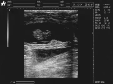
TheWED-2018V full digital veterinary ultrasound scanneris a high-definition convex and linear scanning ultrasound diagnostic system. It is suitable to ultrasound examination and GA measurement of cat,dog, swine, sheep and equine.
It combines microprocessor control and digital scan converter (DSC), and adoptsDigital Beam Forming (DBF), Real-time Dynamic Aperture (RDA) Imaging, Real-timeDynamic Sound Speed Apodization(DRA), Real-time Point by Point Dynamic Receive Focusing (DRF), Digital DynamicFrequency Scanning (DFS), 8-LED Digital TGC, Frame Correlation technology toprovide clear, stable and high-definition images.
Functions
1. The full digital veterinary ultrasound scanner has 6 display modes namely B,B B, B M, B M/M, M, and 4B, and has 256 levels of gray scale.
2. The images can be displayed in real time, and easilyfroze, stored, and loaded.
3. You can even zoom to particular area and up/downconversion, left/right conversion, black/white conversion, and high-capacitymovie playback are all available.
4. Multi-level scanning depth, scanning angel, dynamicrange, sound output power, frame correlation coefficient, number, intervals,and positions of focuses can all be adjusted.
5. Time and date are displayed. You can alsoput the name, genre, age, doctor, and hospital in the annotation.
6. The full digital veterinaryultrasound scanner can measure distance, perimeter, area, and volume,and the gestational age of pigs, horses, cows, sheep, cats, and dogs.
Configurations
The full digital veterinary ultrasound scanner comes with several optional probes to meet the needs in clinicaldiagnosis. It is equipped with PAL-D video output function to take theadvantage of external video printers and large screen displays. High-speedUSB2.0 interface is provided to upload the ultrasound images in real time.
Cased in portable plastic structure, the veterinary ultrasound scanner iscomposed of host machine, transducer (probe), and adapter, and comes withswitching supply without power frequency transformer. Due to the programmabledevices and SMT widely adopted, the integration degree of the whole machine hasbeen greatly improved.
Technical Specification of WED-2018V Veterinary Ultrasound Scanner
| Scanning Mode | Convex/linear/Micro-convex |
| Cine-loop | ≥ 4 00frames |
| Battery Capability | ≥ 4400mAh |
| Standard Configuration | 6.5MHz Vet Rectal Linear Probe |
| Optional Configuration | 3.5MHz Convex Probe |
|
| 7.5MHz HF Linear Probe |
|
| 5.0MHz Micro-Convex Probe |
|
| Video printer |
|
| Trolley |
|
| Car charger |
| Monitor | 10.4 inches |
| Image Storage | ≥ 64 frames |
| Scanning Depth | 240mm( MAX), 16-level adjustable |
| Display Angle | Visual and adjustable |
| Display Mode | B, B B, B M, B 2M, M, 4B |
| Operation Interface | Chinese/ English switchable |
| TGC | Near field, far field, total gain |
| Image Flip | Up/ down, left/ right, black/ white |
| Focus | Focus number, focal span, focal position |
| Image control | Dynamic range, scanning line density Frame correlation, M Speed, Acoustic power |
| Image Process | Image Smoothen/ sharpen,, THI, gamma correction, Histogram, Pseudo Color |
| Real-time depth | 16-level adjustable , Zoom |
| Measurement | Distance, circumference, area, volume, obstetrics( GA for equine, bovine, sheep, swine, cat, dog) |
| Report | Measurement reports automatically generate |
| Body Marks | ≥ 16 types |
| Notation | Date, time, name, Patient ID, sex, age, doctor, hospital full screen words edit, body mark, position indicator |
| Port | Video, XGA, USB2.0 |
WED-2018V
Multi-frequencyProbes
3.5MHzConvex Probe
5.0MHzMicro-convex Probe
6.5MHz Vet Rectal Probe
7.5MHzLinear Probe
1
2
3
4
5
Links:https://www.globefindpro.com/products/90661.html
-
 Bridge Middle Block Cutter
-
 Aroma Card
-
 SKJ-80M Gang Saw Machine
-
 Hydraulic Bridge Cutting Machine
-
 SKJ-80 Gang Saw Machine
-
 CNC intersection Plasma and Flame Pipe Cutting Machine
-
 WED-2000V Veterinary Ultrasound Scanner
-
 Single Phase DC Inverter MMA/TIG Welding Machine
-
 RFID Tag
-
 Manual Stone Polishing Machine
-
 SKJ-100M Gang Saw Machine
-
 Gantry Pillars Cutting Machine (Horizontal Blade)
