FDC8000 Color Doppler Ultrasound System
FDC8000Color Doppler Ultrasound System
Brief Introduction
Combiningimage quality with the versatility of applications, the color Dopplerultrasound system can meet the needs of multiple clinical settings. With itsadvanced detail resolution and blood flow imaging capability, it can be easilydistinguished between cystic and solid structures. It delivers a betterperformance in diagnoses of thyroid, breasts and ovaries.
In addition, the enhanced detail and contrast resolution help identify even themost challenging pathologies. These capabilities help to improve doctor’sability and bring earlier diagnosis. Apart from visualizing vessels that havemultiple velocities per minute in the kidney, this system makes easier for toscan patients from the NICU to geriatrics.
Features
Experience-based Workflow
The experience-based workflow protocolincreases reproducibility and consistency of an echo exam with the colorDoppler ultrasound system.
It uses an expert database of real clinical cases to help recognize differentpositions of the diseases. Randomly choices for measurement labeling functionof vector graphics can be done and refresh the results automatically byadjusting the measuring point, or you can delete any measured value.
Besides, the report page under multiple diagnostic modes can be easily editedbefore printing.
Innovative Technologies
The advanced structure of the color Doppler ultrasound system adoptsbreakthrough technologies coupled with high-rate date acquisition modules andhigh quality image technology delivering excellent image resolution.
All-directionalFree Hands
Due to its all-directional free hands, the monitor can be adjusted to any angleand direction.
15-inchHigh-Resolution Medical LCD
In addition, the 15 inches high-resolution flat screen monitor reduces eyefatigue and improves visibility and clarity.
Enhanced UserInterface and Ergonomic Panel
Besides, enhanced user interface and ergonomic panel reducekeystrokes and manipulations when transforming B, BB, M, CD, PWD, CWD and DirPwr/Pwr mode displays.
Technical Specifications of the Color Doppler Ultrasound System
| Applications | Abdominal, OB/GYN, urology, cardiology, vessel, musculoskeletal, small parts |
| Display modes | B, BB, M, CD, PWD, CWD, DirPwr/Pwr |
| Continuous wave Doppler (CWD) | Expand hemodynamic diagnosis to professional cardiac application, especially congenital heart disease, offering more clinical accuracy and reliability |
| Triplex display | Real-time triplex display B/Color Doppler/Pulsed Spectral Doppler (Three TGCs can be adjusted respectively) |
| Standard transducer | Convex 5C2A (2.0-5.0 MHz) |
| Optional transducer | Linear 12L5A (5.0-12.0 MHz), Transvaginal 8EC4A (4.0-8.0 MHz), Phased array 4V2S (2.0-4.0 MHz) |
| High-precision full digital image technology | Digital beam-forming/real-time continuous dynamic focusing/digital dynamic aperture/real-time dynamic apodization /real-time independent frequency conversion/adjustable focus position |
| High sensitivity blood image (HSI) | Synchronous, homogeneous, clear, sufficient colorful blood image |
| High-definition frequency spectrum image (HDI) | Recognizable sharp marginal blood frequency spectrum echo image |
| Trapezoid imaging (TI) | Offer trapezoidal field of vision to enlarge the view angle of linear imaging |
| Sector expanding image | Offer a wider view of the organs |
| Advanced image processing technology | Automatic image optimizing enhancement, smoothing filter/frame correlation/sharpen/pseudo-color/color priority/grey level transformation/real-time zoom |
| Tissue harmonic imaging (THI) | Suppress speckle noise, brighten image and improve the image quality |
| Automatically-optimized parameters | Adjust the parameters automatically according to the specific clinical diagnostic position so as to save the operating time consumption |
| Powerful measurement software | Versatile clinic-oriented measuring software package |
| i -Station | Versatile image format, such as AVI, JPG, TIF, BMP, SEQ, DICOM3.0 and super high medical report capacity storage, fast report management, such as preview, edit, print and transmission |
| Port | USB2.0, RS-232, XGA, DICOM3.0, PAL-D, Network, DVD-R/W |
| Annotation | Text, body marks, general notes |
| Report | Automatic report generation |
| Monitor | 15 inches medical LCD |
| Panel | Ergonomic panel |
Transducers
1
2
3
4
Quality images
Cardiac imaging in Doppler color-mode
Carotid Bifurcation
Frequency Spectral Doppler of Regurgitation
Real-time triplex display BColor Doppler Pulsed Spectral Doppler
Reflux Dilated Long SaphenousVein
Thyroid imaging in color mode
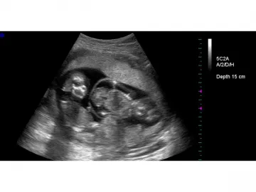
Twins imaging in 2D-mode
Vaginal imaging in color Doppler mode
FDC8000 Instruction Manual
FDC8000B/BB/M/CD/PWD/CWD/DirPwr/Pwr,。(color doppler ultrasound scanner),、、。(ultrasoundmachine),,。,。
(color doppler ultrasound scanner)CE,,。、、。、、,。
Links:https://globefindpro.com/products/90686.html
-
 WED-M1V Veterinary Ultrasound Scanner
-
 3 Phase DC Inverter CO2/Metal Active Gas Welding Machine
-
 Gantry CNC Plasma and Flame Cutting Machine
-
 Amusement Rides, Seesaw
-
 Brazing Furnace
-
 Safety Floor Mat
-
 Member Card
-
 Bridge Multi-blade Block Cutter
-
 Irregular Shape Card
-
 Contactless IC Card 13.56MHz
-
 Stone Cutting Machine
-
 Automatic Bridge Cutting Machine
